



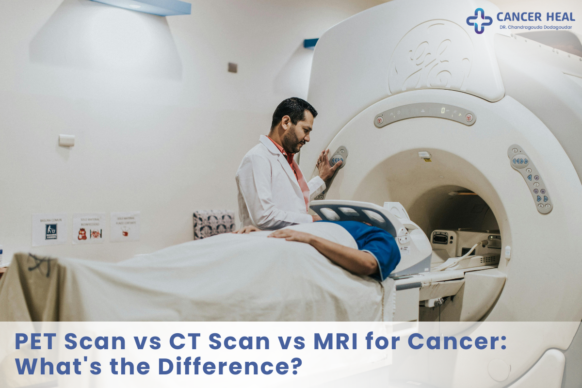
When facing a potential oncology diagnosis or evaluating treatment progress, selecting the appropriate diagnostic scan is critical. Navigating the choices between a PET scan vs CT scan vs MRI for cancer can feel overwhelming, but each imaging technology serves a unique, highly specialized role. CT scans capture anatomical cross-sections, MRIs provide detailed soft-tissue resolution, and PET scans track metabolic cellular activity. Understanding how these scans compare helps patients and families make informed medical decisions.
In this comprehensive guide, you will learn the fundamental differences between these three imaging modalities, how doctors determine the best scan for specific tumor types, how imaging improves diagnostic accuracy, and the top imaging packages available today.
What Is Cancer Diagnostic Imaging?
Cancer diagnostic imaging refers to specialized non-invasive scanning technologies used to detect, locate, stage, and monitor malignant growths inside the human body. By utilizing X-rays, magnetic fields, or radioactive tracers, these modalities create detailed visual representations of internal organs, bones, and metabolic functions to guide precision oncology treatment.
Each imaging modality provides distinct clinical insights. Computed Tomography (CT) uses rotating X-rays to generate 3D anatomical slices. Magnetic Resonance Imaging (MRI) uses strong magnetic fields and radio waves to map soft tissues without radiation. Positron Emission Tomography (PET) injects a safe radiotracer—most commonly fluorodeoxyglucose (FDG)—to highlight hyperactive cancer cells consuming sugar at elevated rates.
Key Components of Diagnostic Scans
-
Computed Tomography (CT): High-speed X-ray cross-sections optimized for dense structures, lungs, abdominal organs, and bone details.
-
Magnetic Resonance Imaging (MRI): Radiofrequency and magnetic mapping delivering high-contrast images of soft tissues, brain, spinal cord, and pelvic organs.
-
Positron Emission Tomography (PET): Metabolic molecular imaging that detects abnormal cellular energy consumption before physical structural changes appear.
-
Hybrid Imaging (PET-CT): Combined technology merging metabolic PET data with anatomical CT precision in a single scanning session.
Why Is Choosing the Right Cancer Scan Important?
Rising cancer cases in India highlight the vital role of precise early imaging in modern healthcare. According to recent public health data, the overall cancer incidence in India continues to rise, with lung, breast, cervical, and oral malignancies ranking among the most common cancers in India. Catching tumors early directly impacts treatment success and helps lower the overall cancer death rate in India.
Selecting the wrong scan can delay accurate staging or lead to inconclusive results. For example, a CT scan might reveal an enlarged lymph node, but a PET scan determines whether that node contains active, proliferating cancer cells. Conversely, an MRI is far superior for visualizing brain or spinal tumors where bone artifacts distort CT images. Utilizing the appropriate imaging modality ensures timely, targeted oncological interventions.
How Does PET Scan vs CT Scan vs MRI for Cancer Work?
Navigating the diagnostic imaging process involves three structured steps designed to ensure maximum image clarity and patient safety.
Step 1: Patient Preparation and Contrast Administration
Depending on the assigned scan, you may need to fast for several hours. For a PET scan, a radiotracer is injected, requiring a 60-minute quiet resting period while the tracer circulates. For CT or MRI scans, oral or intravenous contrast dyes may be administered to highlight blood vessels and organ boundaries.
Step 2: Image Acquisition in the Scanner
You lie comfortably on a motorized table that slides into the scanner. CT scans are extremely fast, taking only 1 to 5 minutes. MRI scans require 20 to 45 minutes inside a magnetic tube while radiofrequency pulses operate. PET scans take approximately 20 to 30 minutes to complete full-body acquisition.
Step 3: Fusion and Radiological Analysis
Subspecialized radiologists and nuclear medicine physicians analyze the digital image datasets. In combined PET-CT systems, advanced software merges metabolic heat maps directly over structural CT slices, pinpointing exact anatomical locations of active tumors.
Top Recommended Diagnostic Imaging Packages & Scans
Choosing an accredited imaging center with high-resolution technology guarantees reliable diagnostic reports. Below are top-recommended diagnostic imaging evaluation packages available across leading medical centers.
1. Comprehensive PET-CT Whole-Body Staging Scan
A full-body metabolic and structural evaluation designed for accurate cancer staging, metastasis evaluation, and therapy response monitoring.
-
Included Technologies: Whole-body FDG PET scan integrated with multi-slice contrast-enhanced CT.
-
Best For: Staging lung, breast, lymphoma, colorectal, and esophageal malignancies.
2. High-Resolution Brain & Spine MRI Package
A specialized soft-tissue imaging package designed to evaluate central nervous system tumors with high diagnostic precision.
-
Included Technologies: Multi-planar T1, T2, FLAIR, and Contrast-Enhanced MRI sequences.
-
Best For: Detecting brain metastases, primary glioblastomas, spinal cord lesions, and soft-tissue sarcomas.
3. Multi-Slice Chest & Abdomen Diagnostic CT Package
An ultra-fast anatomical imaging package providing detailed cross-sectional views of major thoracic and abdominal organs.
-
Included Technologies: High-definition IV contrast-enhanced Computed Tomography.
-
Best For: Evaluating pulmonary nodules, liver lesions, renal masses, and pancreatic tumors.
4. Specialized Pelvic MRI Package for Women & Men
A targeted soft-tissue scan offering detailed anatomical visualization of pelvic organs.
-
Included Technologies: Diffusion-Weighted Imaging (DWI) and dynamic contrast-enhanced pelvic MRI.
-
Best For: Staging prostate, uterine, cervical, and rectal cancers.
Benefits of Modern Cancer Imaging Modalities
Precise Cancer Staging
-
What it is: Determining the exact size, depth, and spread of a tumor throughout the body.
-
Why it matters: Prevents unnecessary surgeries when cancer has already spread and guides precise radiation therapy.
-
Expected outcome: Highly tailored, stage-specific treatment plans.
Treatment Efficacy Evaluation
-
What it is: Assessing whether chemotherapy or radiation is successfully killing tumor cells.
-
Why it matters: PET scans show reduced metabolic activity weeks before a physical tumor shrinks on standard structural scans.
-
Expected outcome: Ability to switch ineffective therapies early, saving time and medical expenses.
High-Resolution Target Mapping
-
What it is: Generating exact 3D anatomical coordinates for radiation oncologists and surgical teams.
-
Why it matters: Allows surgeons to preserve healthy surrounding tissues and critical nerves.
-
Expected outcome: Safer surgical interventions and reduced treatment-related side effects.
Challenges and Common Mistakes in Cancer Imaging
Myth: “A PET Scan Can Replace All CT and MRI Scans”
-
Problem: Assuming a PET scan is always the superior choice for every type of cancer.
-
Cause: Overestimating the standalone capability of metabolic imaging without considering spatial resolution limits.
-
Solution: Understand that PET scans lack fine anatomical detail and are almost always combined with CT or MRI for precision.
Myth: “MRIs Are Dangerous Because of High Radiation Levels”
-
Problem: Avoiding necessary MRI procedures out of fear of radiation exposure.
-
Cause: Confusing MRI technology with ionizing radiation used in CT scans and X-rays.
-
Solution: Recognize that MRI uses powerful magnetic fields and radio waves, producing zero ionizing radiation.
Myth: “Every Lump Seen on a CT Scan Is Cancer”
-
Problem: Panicking over non-cancerous anatomical abnormalities reported on imaging scans.
-
Cause: Inability of structural scans alone to distinguish benign cysts, scar tissue, or inflammation from active malignancies.
-
Solution: Correlate structural findings with PET metabolic imaging or tissue biopsies for definitive diagnosis.
8 Actionable Best Practices for Cancer Diagnostic Scans
-
Follow Fasting Instructions Strictly: For PET scans, high blood glucose levels block radiotracer absorption, leading to inaccurate results.
-
Disclose Metal Implants Before MRI: Inform your radiologist about pacemakers, joint replacements, or metallic fragments due to strong magnetic fields.
-
Check Kidney Function Prior to Contrast: Ensure blood creatinine levels are tested before receiving contrast dyes for CT or MRI scans.
-
Inform Your Doctor About Claustrophobia: Request mild sedation or opt for wide-bore MRI machines if confined spaces cause anxiety.
-
Avoid Strenuous Exercise Before PET Scans: Muscle exertion causes radiotracer uptake in healthy muscle tissue, mimicking tumor activity.
-
Bring Previous Scan Records: Always provide past imaging CDs and reports so radiologists can compare tumor size changes over time.
-
Request Low-Dose Protocols When Possible: For routine CT follow-ups, ask if low-dose CT protocols are suitable for your condition.
-
Select Accredited Imaging Centers: Ensure the facility utilizes modern 3T MRI machines and digital PET-CT scanners for maximum image resolution.
When to Choose PET Scan vs CT Scan vs MRI for Cancer
Selecting between these modalities depends on the primary organ involved and the medical question being asked. CT scans serve as the workhorse for initial chest and abdominal evaluations. MRIs are the gold standard for brain, spinal, bone, and soft-tissue soft-tissue detail. PET scans excel at whole-body metabolic staging, detecting hidden metastases, and evaluating treatment response.
Comparison Table: Cancer Staging Scans Compared
| Feature | Computed Tomography (CT) | Magnetic Resonance Imaging (MRI) | Positron Emission Tomography (PET) |
| Primary Mechanism | Rotating X-ray beams | Powerful magnets & radio waves | Radiotracer glucose uptake |
| Primary Strength | Fast 3D anatomical images of bones & lungs | Superior soft-tissue resolution without radiation | Maps cellular metabolic activity |
| Radiation Exposure | Low to moderate ionizing radiation | None (Zero radiation) | Low radioactive tracer exposure |
| Scan Duration | 1 to 5 minutes | 20 to 45 minutes | 20 to 30 minutes (plus prep time) |
| Best Clinical Use | Lung nodules, liver/kidney masses, emergency staging | Brain, spine, pelvic organs, soft-tissue sarcomas | Recurrence checking, metastasis, therapy tracking |
| Average Cost Range (India) | ₹3,000 – ₹8,000 | ₹6,000 – ₹15,000 | ₹15,000 – ₹35,000 |
Frequently Asked Questions
What is the best imaging test for cancer detection?
There is no single best imaging test for all cancers. CT scans excel at detecting chest, lung, and abdominal tumors quickly. MRI is superior for brain, spinal cord, and pelvic soft tissues. PET scans are best for evaluating whole-body metabolic activity, detecting distant metastases, and verifying whether a tumor is actively growing or responding to chemotherapy treatments.
What is the main difference between PET CT and MRI scan?
The primary difference between PET CT and MRI scan lies in what they measure. An MRI provides detailed anatomical structural images of soft tissues using magnetic fields without radiation. A PET-CT combines structural X-ray details with metabolic radiotracer tracking, showing both what a tumor looks like and how active its cellular metabolism is.
How does MRI vs CT scan for cancer diagnosis compare?
In comparing MRI vs CT scan for cancer diagnosis, CT scans use X-rays to generate rapid images of bones, lungs, and abdominal organs. MRI uses magnetic fields to produce higher-contrast soft-tissue resolution without radiation, making MRI far better for brain, spinal cord, muscle, and pelvic organ evaluations where subtle tissue variations matter.
What is the PET scan accuracy for cancer staging?
The PET scan accuracy for cancer staging is exceptionally high, often exceeding 85% to 95% for malignancies like lymphoma, lung cancer, and melanoma. By evaluating metabolic cellular activity throughout the entire body, PET scans detect microscopic metastases in lymph nodes and distant organs long before structural size changes become visible on standard CT or MRI scans.
Why do doctors order a combined PET-CT scan instead of just a CT scan?
A standard CT scan shows tumor shape and size, but cannot distinguish between active cancer tissue and inactive scar tissue. Adding PET metabolic data allows doctors to pinpoint exact areas of active cellular proliferation mapped directly onto anatomical CT slices, providing unmatched diagnostic clarity for cancer staging and treatment planning.
How do cancer staging scans compared help reduce the cancer death rate in India?
Comparing cancer staging scans helps oncologists select the right scan immediately, avoiding diagnostic delays. Accurate staging ensures patients receive stage-appropriate treatments—such as localized surgery for early stages or targeted systemic therapy for advanced stages—significantly improving survival rates and reducing overall mortality across oncology centers in India.
Are PET scans safe regarding radiation exposure?
Yes, PET scans are safe. The radioactive tracer used (such as FDG) carries a low radiation dose comparable to a standard diagnostic CT scan. The tracer decays rapidly and leaves the body naturally through urine within several hours. Patients are advised to drink plenty of water after the scan to flush out residual tracer.
Which scan is best for detecting brain tumors or spinal lesions?
MRI is the undisputed gold standard for detecting brain tumors, spinal cord lesions, and soft-tissue sarcomas. Because bones surround the brain and spinal cord, CT scans often produce dense shadows and artifacts. MRI bypasses bone density, delivering crystal-clear contrast between normal brain parenchyma, edema, and tumor boundaries.
Key Takeaways
-
Each Scan Has a Role: CT provides fast structural views, MRI delivers unmatched soft-tissue detail, and PET tracks metabolic tumor activity.
-
Combined Systems Offer Precision: Hybrid PET-CT scans merge structural and functional data for superior staging accuracy.
-
MRI Contains Zero Radiation: Ideal for brain, spine, and pelvic imaging without ionizing radiation risks.
-
PET Assesses Treatment Success: Evaluates whether chemotherapy is working weeks before physical tumor dimensions shrink.
Final Verdict
Understanding how diagnostic scans function removes uncertainty when facing medical evaluations. While CT scans offer rapid anatomical overviews and MRIs capture intricate soft-tissue structures, PET scans reveal active cellular metabolism. Relying on your oncologist’s guidance to select the right scan—or a combination of modalities—ensures accurate staging, optimal treatment selection, and better long-term health outcomes.
Conclusion
Comparing a PET scan vs CT scan vs MRI for cancer reveals that these technologies are complementary tools rather than competing choices. Reviewing key cancer facts in India highlights the immense value of timely diagnostic imaging in turning complex diagnoses into manageable conditions. By consulting with experienced radiologists and oncologists, you can choose the ideal diagnostic scan package for your specific health needs. Take charge of your medical journey today by discussing advanced imaging options with your specialist.
